John Grigg
Senior Lecturer
Department of Ophthalmology, University of Sydney
[email protected]
- Definition:
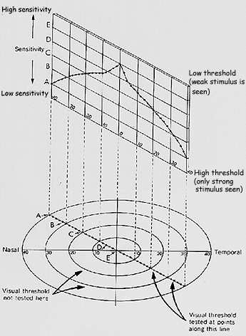
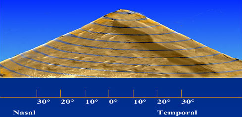
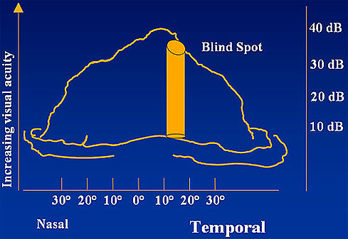
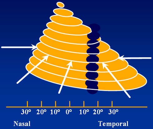
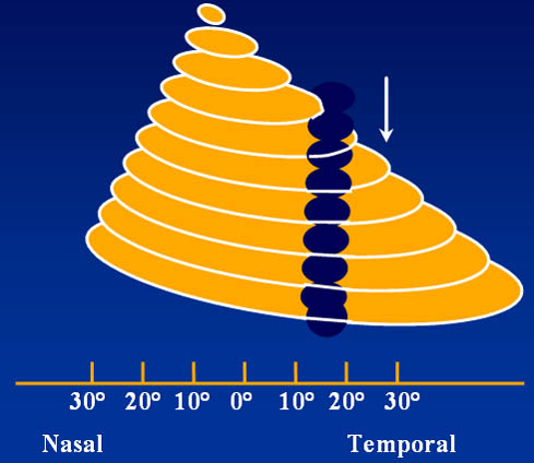
- That portion of space in which objects are simultaneously visible to the steadily fixing eye. - Traquair:
- "an island hill of vision surrounded by a sea of blindness".
- Scotoma
- focal area of decreased sensitivity surrounded by more normal sensitivity an island of partial blindness within the confines of a normal visual field.
- described as:
• absolute or relative
• anatomic eg paracentral
• shape eg ring
- Contraction
- generalised depression with absolute loss of some portions of the outermost border to the most intense stimulus
- area of defective visual field that is totally blind to all stimuli.
- Depression
- an area of decreased sensitivity without a more normal surround
• constriction of isopters
• general or local
- Stimulus variables
- Background illumination
- Observer variables
- Confrontation
- Tangent
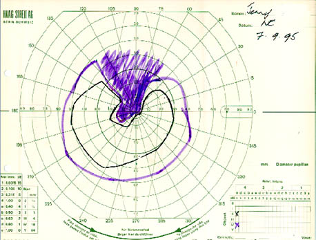
- Goldmann
- Amsler Grid
- Computerized perimetry
- Angular size of the target
- VA decreases with increasing eccentricity; 6/12 at 3°, 6/60 at 20° and 6/120 at 40°
- Larger target sizes needed for more peripheral areas of the retina - Temporal characteristics of the target
- Duration, movement of stimulus, background luminance - Colour of the target
- Provide greater sensitivity than white (this may be due to stimulating a larger area of the retina)
- Pupillary diameter
- Ocular media
- Lids
- Refraction
- unnecessary for 30° - 60° fields. - Visual Pathway pathology
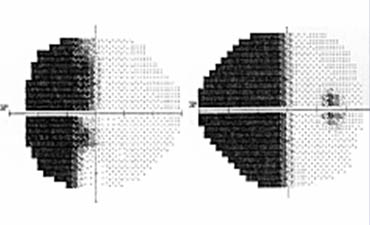
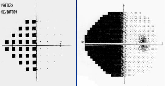
- Supratentorial
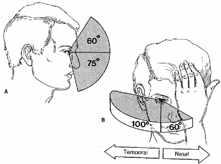
- Greatest use within 30 degrees
- Screen should be 2 meters square
- Markings - radial marks are 30 degrees apart
- Concentric circles marked at 5,10,15,20 degrees for the 2 meter distance (these will be 10,20,30, & 40 degrees at 1 meter)
- Fixation targets range of sizes from 1mm to 10cm
- Patient seated at the appropriate distance from the screen
- Size of the fixation target depends on the patient's refractive error, the size of the scotoma that is suspected or being elicited
- Begin by outlining the blind spot
- Mark disappearance or reappearance with a black pin
- Move from blind to seeing areas
- Define upper and lower sensitivity limits
- use a small target to detect an early constriction of inner isopters
- use as large a target as possible to demonstrate true nature of field defect
- Use red target to check findings
- Neurological field defects best detected in the central 30 degrees
- Glaucoma rare to find defects in the far periphery unless the central field is also disturbed
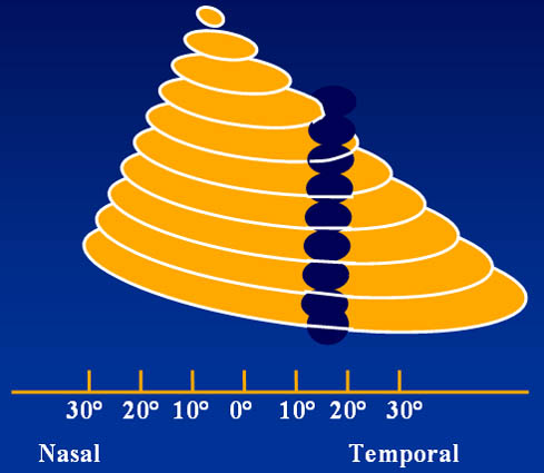
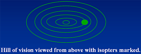
- Method of indicating a flat projection of a hemispherical surface
- Recording a survey of the hill of vision in terms of contour lines
- Therefore the field is recorded as a series of irregular circles
- Recorded as the patient sees the screen
- Record the target size and the distance from the screen e.g. 3/2000
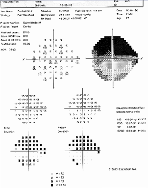
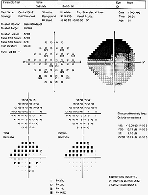
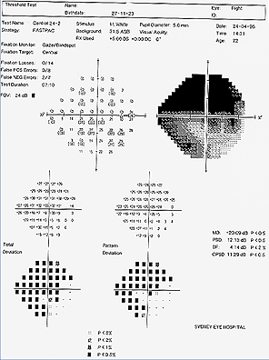
- Generalised depression of field
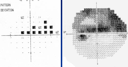
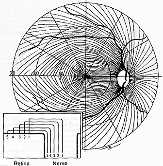
- Localised nerve fibre bundle defects/paracentral changes
- Typical arcuate scotomata
- Nasal step (central or peripheral), temporal sector defect
- Advanced constriction (central and temporal island only)
- Not enlarged blind spot - as due to peripapillary atrophy
- In normal tension glaucoma field defects may be closer to fixation and steeper edge scotomas
- Defects often in superior field first
Goldmann field
