Dr Alex Hunyor
Vitreoretinal Unit
Sydney Eye Hospital
- How to turn on direct ophthalmoscope
- Rheostat
- Filters, reticle
- Dioptre adjustment
- Examination technique
• right eye/hand/eye
• fix in distance
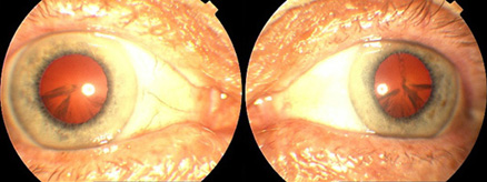
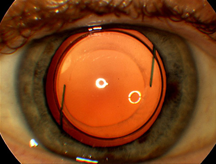
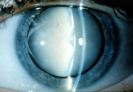
• red reflex
- Landmarks
• follow retinal vessels to disc
• veins darker and thicker
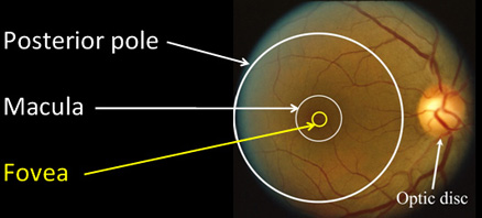
• macula temporal (lateral) to disc - Normal Findings
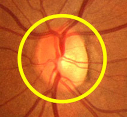
• optic disc
• retinal vessels
• macula

- Optic disc
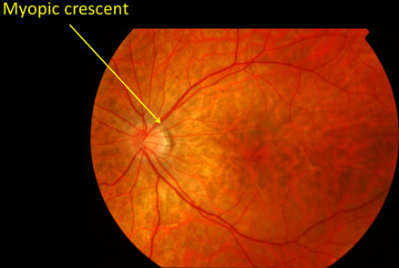
• Colour: optic cup vs neuroretinal rim
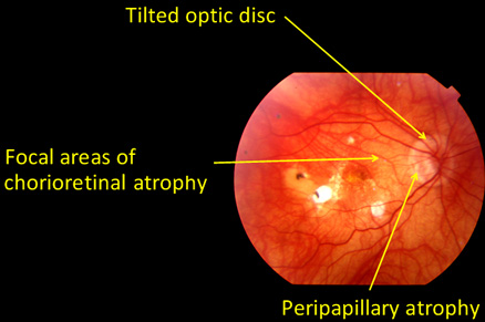
• Shape: round, oval, tilted
• Margins: sharp, blurred
• Cup/Disc ratio: normal range, symmetry
-
• Colour: pink
• Shape: sl vertical oval
• Margins: well defined
• Optic cup: depressed central area
• Look at retinal vessels going over edge of cup
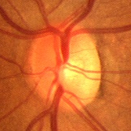
• Cup/Disc ratio 0.3

- Retinal Blood Vessels
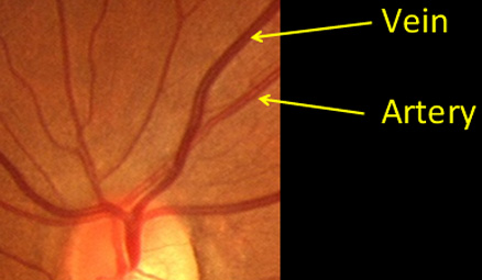
• Veins are wider and darker
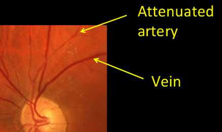
• Normal ratio of calibre of arteries to veins (A-V ratio) is 3:2
• May be abnormal due to narrowing of arteries or dilation of veins
• May observe changes at arteriovenous crossings (A-V nipping)
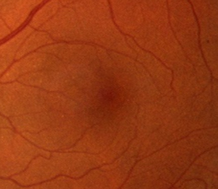
- Macula
• Centre of macula approx 2 disc diameters from edge of the optic disc and slightly lower
• No retinal blood vessels in centre of macula
• Foveal light reflex in younger people
-
• No foveal reflex in this photo
• Darker pigment
• Absent retinal vessels in fovea (centre of macula)
